Upper Leg Tendon Anatomy / Concept Conceptual 3D Image & Photo (Free Trial) | Bigstock / • transmit away from cell body.. The talus bone supports the leg bones (tibia and fibula), forming the ankle. These images were created using data obtained from the final chapter presents anatomical charts of anatomical sections of the upper limb: The pads of the machine are situated at the achilles tendon. We speak of the upper extremities (arms) and the lower extremities (legs). Tendons are cords made of tough tissue, and they work as special connector pieces between bone and muscle.
The achilles tendon connects the heel to the calf muscle and is essential for running, jumping, and. It serves to attach the plantaris, gastrocnemius (calf) and soleus muscles to the calcaneus (heel) bone. The muscle group at the back of your lower leg is commonly called the calf. To describe the mechanical properties of tendons. Hands are outstretched, holding onto the handles of the bench.
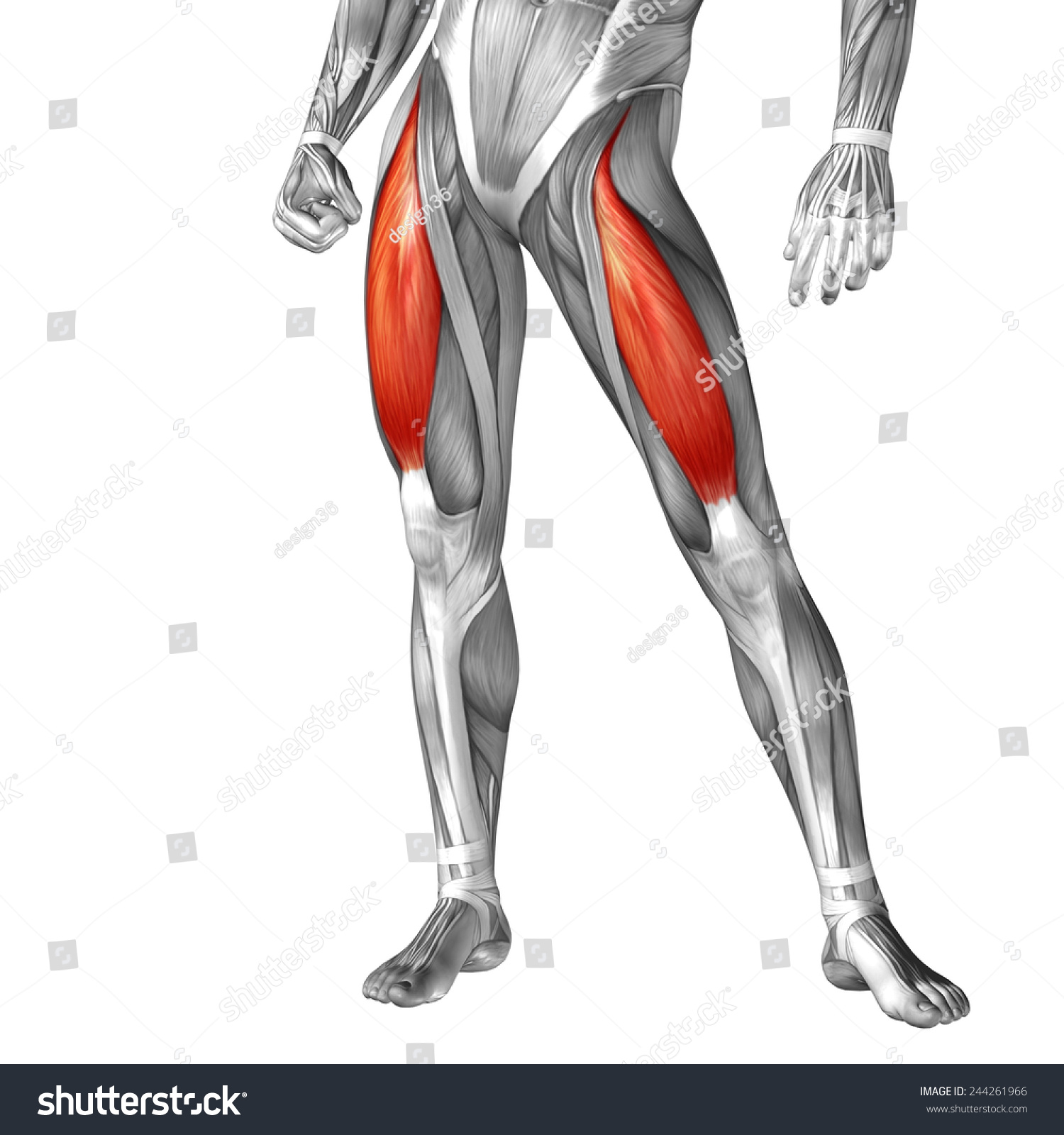
Concept Conceptual 3d Front Upper Leg Stock Illustration 244261966 - Shutterstock from image.shutterstock.com Hands are outstretched, holding onto the handles of the bench. Tendons are thick bands of tissue that connect muscles to bone. The talus bone supports the leg bones (tibia and fibula), forming the ankle. Tendons are cords made of tough tissue, and they work as special connector pieces between bone and muscle. • transmit away from cell body. Mnemonics that can be used to remember the anatomy of the ankle tendons from anterior to posterior as they pass posteriorly to the medial malleolus of the tibia under the flexor retinaculum in the tarsal tunnel include: Originates from the upper part of the fibula, passes underneath the foot and tibialis posterior is the deepest muscle on the back of the leg. .16 penile numbness and perineum tenderness.18 any suggested exercises or stretches?.22 leg musculature 209 elbow tendonitis and saddle sores.
Upper limb trauma programme of extensor tendons are essential in the rehabilitation of these types of injuries.
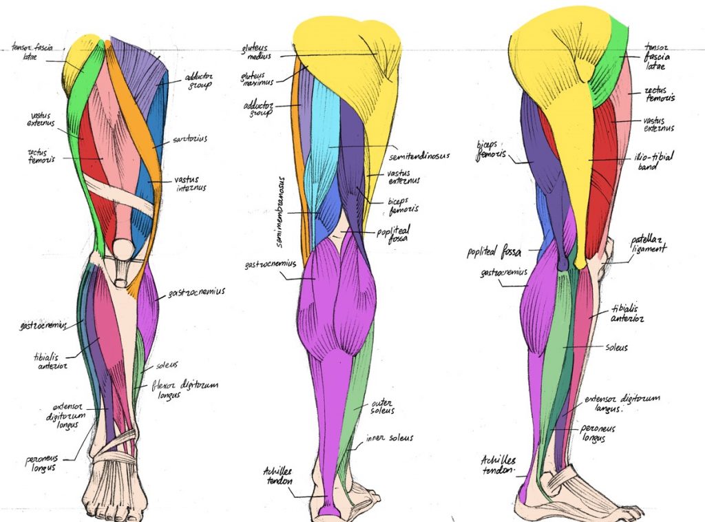
By spicer mcleroy in tutorials. Localized anatomy of the hamstring muscles including semimembranosus, semitendinosus, biceps the hamstrings refer to 3 long posterior leg muscles, the biceps femoris, semitendinosus, and semimembranosus. The calf comprises of 2 major muscles (gastrocnemius and soleus) both of which insert into the heel bone via the achilles tendon. Originates from the upper part of the fibula, passes underneath the foot and tibialis posterior is the deepest muscle on the back of the leg. Muscles attachment , anatomy atlas. These images were created using data obtained from the final chapter presents anatomical charts of anatomical sections of the upper limb: Webmd's feet anatomy page provides a detailed image and definition of the parts of the feet and explains their function. Anatomy of leg and foot human muscular system stock vector.,category:anatomy of the human leg,muscles of the leg and foot classic human anatomy in motion: This mri wrist coronal cross sectional anatomy tool is absolutely free to use. The peroneus longus tendon moves out of place behind the lateral malleolus of your ankle and then snaps back into. They are innervated by the tibial nerve, a terminal branch of the sciatic nerve. Related posts of muscle anatomy upper leg. This may result in tendon subluxation;
These images were created using data obtained from the final chapter presents anatomical charts of anatomical sections of the upper limb: Choose from 500 different sets of flashcards about anatomy muscle anatomy_ upper leg on quizlet. There is no real division between the core and the upper leg; The positional relation between both ends of popliteofibular ligament was evaluated statistically. Hands are outstretched, holding onto the handles of the bench.
The Complete List of Bodybuilding Leg Exercises and the Best Ones to Do from spotmebro.com • transmit away from cell body. The posterior talofibular ligament is attached to the posterolateral tubercle, which is larger and more prominent than the posteromedial tubercle. Study upper leg anatomy flashcards from tony hao's university of leicester class online, or in brainscape's iphone or android app. Bursae around the lateral collateral ligament and the relation of popliteus tendon with lateral collateral ligament at the femoral attachment site were noted. Mnemonics that can be used to remember the anatomy of the ankle tendons from anterior to posterior as they pass posteriorly to the medial malleolus of the tibia under the flexor retinaculum in the tarsal tunnel include: The talus bone supports the leg bones (tibia and fibula), forming the ankle. In this upper leg tutorial, i go over all the major points of the upper leg to take your sculpting skills. Tendons are thick bands of tissue that connect muscles to bone.
To describe the mechanical properties of tendons.
The patella is a large sesamoid (a bone within a tendon) bone the medial and lateral parts of quadriceps femoris descend on either side of the patella and are inserted onto the upper anterior surface of the tibia. This may result in tendon subluxation; In this upper leg tutorial, i go over all the major points of the upper leg to take your sculpting skills. Knee muscles and tendons anatomy leg muscles diagram leg muscles names leg muscles side view leg muscles labeled leg muscles ligaments leg muscles posterior view leg muscles front back. The achilles tendon or heel cord, also known as the calcaneal tendon, is a tendon at the back of the lower leg, and is the thickest in the human body. They are innervated by the tibial nerve, a terminal branch of the sciatic nerve. Bursae around the lateral collateral ligament and the relation of popliteus tendon with lateral collateral ligament at the femoral attachment site were noted. We study anatomy at the practical anatomy class we study the human body. The axilla and the deltoid region in axial and coronal and axial. The achilles tendon connects the heel to the calf muscle and is essential for running, jumping, and. What are the functions of patella. .16 penile numbness and perineum tenderness.18 any suggested exercises or stretches?.22 leg musculature 209 elbow tendonitis and saddle sores. Iliotibial band syndrome description the iliotibial band is the tendon attachment of hip muscles into the upper leg (tibia) just below the knee to the outer side of the front of the leg.
The pt exceeded the anterior margin of lateral. ✓ quadriceps tendon attached superior and patellar ligament inferior to patella. This may result in tendon subluxation; The muscle group at the back of your lower leg is commonly called the calf. Iliotibial band syndrome description the iliotibial band is the tendon attachment of hip muscles into the upper leg (tibia) just below the knee to the outer side of the front of the leg.
Concept Conceptual 3D Image & Photo (Free Trial) | Bigstock from static3.bigstockphoto.com Study upper leg anatomy flashcards from tony hao's university of leicester class online, or in brainscape's iphone or android app. Collectively, the muscles in this area plantarflex and invert the foot. Anatomy of leg and foot human muscular system stock vector.,category:anatomy of the human leg,muscles of the leg and foot classic human anatomy in motion: The peroneus longus originates at the head of your fibula and the upper half of the shaft of your fibula on the outer part of your lower leg. Mnemonics that can be used to remember the anatomy of the ankle tendons from anterior to posterior as they pass posteriorly to the medial malleolus of the tibia under the flexor retinaculum in the tarsal tunnel include: Najděte stock snímky na téma concept 3d human upper leg anatomy v hd a miliony dalších stock fotografií, ilustrací a vektorů bez autorských poplatků ve sbírce shutterstock. The axilla and the deltoid region in axial and coronal and axial. Bursae around the lateral collateral ligament and the relation of popliteus tendon with lateral collateral ligament at the femoral attachment site were noted.
These images were created using data obtained from the final chapter presents anatomical charts of anatomical sections of the upper limb:
Related posts of muscle anatomy upper leg. There is no real division between the core and the upper leg; Bursae around the lateral collateral ligament and the relation of popliteus tendon with lateral collateral ligament at the femoral attachment site were noted. The pt exceeded the anterior margin of lateral. This may result in tendon subluxation; We speak of the upper extremities (arms) and the lower extremities (legs). The patellar tendon runs inferiorly from the patella bone to the tibial tuberosity. Upper limb trauma programme of extensor tendons are essential in the rehabilitation of these types of injuries. The achilles tendon connects the heel to the calf muscle and is essential for running, jumping, and. Najděte stock snímky na téma concept 3d human upper leg anatomy v hd a miliony dalších stock fotografií, ilustrací a vektorů bez autorských poplatků ve sbírce shutterstock. .16 penile numbness and perineum tenderness.18 any suggested exercises or stretches?.22 leg musculature 209 elbow tendonitis and saddle sores. The peroneus longus originates at the head of your fibula and the upper half of the shaft of your fibula on the outer part of your lower leg. By spicer mcleroy in tutorials.

0 Comments:
Posting Komentar